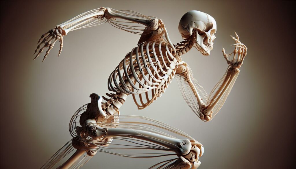
I’ve always been fascinated by the incredible framework that keeps us upright and moving – our skeleton. This remarkable structure consists of 206 bones that work together like a perfectly engineered machine protecting our vital organs and enabling everyday movements.
As a passionate anatomist I’ve studied how the skeletal system serves as more than just a support structure. It’s also a calcium storage facility produces blood cells and acts as a vital anchor point for our muscles. While many think of bones as rigid unchanging structures they’re actually dynamic living tissues that continuously rebuild and repair themselves throughout our lives.
Key Takeaways
- The human skeleton consists of 206 bones that not only provide structural support but also store calcium, produce blood cells, and anchor muscles.
- The skeletal system performs 5 essential functions: supporting body weight, protecting vital organs, producing blood cells, storing minerals, and enabling movement.
- There are 4 main types of bones: long bones (like femur), short bones (wrist bones), flat bones (skull), and irregular bones (vertebrae).
- The skeleton is divided into two main sections: the axial skeleton (80 bones) which forms the central core, and the appendicular skeleton (126 bones) which includes limbs and girdles.
- Bones are dynamic living tissues that continuously remodel themselves, with approximately 10% of total bone mass being replaced annually.
- Common skeletal disorders like osteoporosis affect millions, with 10.2 million Americans age 50+ having the condition and 43.4 million having low bone mass.
Anatomy:buuf4tqlbqo= Skeleton
The human skeletal system forms a complex framework of bones interconnected through joints ligaments. I examine how this remarkable system provides structural support while enabling precise movements through its specialized components.
Basic Functions of the Skeleton
The skeletal system performs 5 essential functions in the human body:
- Supports body weight by creating a rigid internal framework
- Protects vital organs like the brain heart spinal cord through specialized bone structures
- Produces blood cells in red bone marrow located inside specific bones
- Stores minerals including calcium phosphorus for metabolic functions
- Enables movement by providing attachment points for muscles tendons
Types of Bones in the Body
The skeletal system contains 4 distinct bone categories:
| Bone Type | Description | Examples |
|---|---|---|
| Long Bones | Cylindrical shaft with ends | Femur Humerus |
| Short Bones | Cube-shaped with equal dimensions | Wrist Ankle bones |
| Flat Bones | Thin plates with broad surfaces | Skull Sternum |
| Irregular Bones | Complex shapes | Vertebrae Hip bones |
- Long bones contain hollow shafts filled with marrow
- Short bones provide stability flexibility in complex joints
- Flat bones create protective shields around organs
- Irregular bones accommodate specific mechanical functions based on their location
Axial Skeleton Components
The axial skeleton forms the central core of the anatomy:buuf4tqlbqo= skeleton, comprising 80 bones that protect vital organs and provide structural support. These bones create a framework for attachment of the appendicular skeleton and maintain the body’s vertical alignment.
Skull and Cranial Bones
The skull consists of 22 bones: 8 cranial bones and 14 facial bones. The cranial bones include the frontal, parietal (2), temporal (2), occipital, sphenoid and ethmoid bones. These bones fuse together through immovable joints called sutures to form a protective case around the brain. The facial bones shape the face, support sensory organs and create attachment points for muscles used in chewing and expression.
Vertebral Column Structure
The vertebral column contains 33 individual vertebrae arranged in five distinct regions: cervical (7), thoracic (12), lumbar (5), sacral (5 fused) and coccygeal (4 fused). Each vertebra features a body, vertebral arch and processes for muscle attachment. Between most vertebrae lie intervertebral discs that act as shock absorbers and allow spinal flexibility. The spinal canal within the vertebral column houses and protects the spinal cord.
Rib Cage and Sternum
The rib cage encompasses 12 pairs of ribs, the sternum and thoracic vertebrae. The ribs connect to the thoracic vertebrae posteriorly and attach to the sternum anteriorly through costal cartilage. The sternum consists of three parts: manubrium, body and xiphoid process. This cage-like structure protects vital organs including the heart, lungs and major blood vessels while allowing for respiratory movements through the action of intercostal muscles.
Appendicular Skeleton Elements
The appendicular skeleton consists of 126 bones that form the upper and lower extremities plus the shoulder and pelvic girdles. These bones enable precise movements ranging from fine motor control in the fingers to powerful locomotion in the legs.
Upper Extremity Bones
The upper extremity contains 32 bones in each arm connected to the axial skeleton through the shoulder girdle. Here’s the breakdown of bones:
- Shoulder girdle (2 per side):
- Clavicle (collarbone)
- Scapula (shoulder blade)
- Arm and forearm (3 bones):
- Humerus in upper arm
- Radius on thumb side
- Ulna on pinky side
- Hand (27 bones):
- 8 carpals in the wrist
- 5 metacarpals forming the palm
- 14 phalanges creating fingers (2 in thumb 3 in other digits)
- Pelvic girdle:
- Hip bone (fusion of ilium ischium pubis)
- Leg bones (3 major bones):
- Femur (thighbone)
- Tibia (shinbone)
- Fibula (smaller leg bone)
- Foot (26 bones):
- 7 tarsals forming ankle and heel
- 5 metatarsals creating the foot arch
- 14 phalanges forming toes (2 in big toe 3 in other toes)
| Region | Number of Bones | Key Functions |
|---|---|---|
| Upper Extremity (each) | 32 | Fine motor control manipulation |
| Lower Extremity (each) | 31 | Weight-bearing locomotion |
| Total Appendicular | 126 | Movement stability support |
Bone Growth and Development
Bone development follows distinct stages from embryonic formation through adulthood, involving complex cellular processes and hormonal regulation. The human skeletal system transforms from flexible cartilage to rigid bone through specialized mechanisms.
Ossification Process
Ossification transforms soft tissue into hardened bone through two primary methods: intramembranous ossification in flat bones like the skull and endochondral ossification in long bones like the femur. Primary ossification centers appear in the diaphysis during fetal development at 8 weeks, while secondary centers emerge in the epiphyses after birth. This process requires:
- Osteoblasts depositing calcium phosphate crystals into collagen fibers
- Blood vessel formation to supply nutrients
- Mineralization of the bone matrix
- Growth plate activity between the diaphysis and epiphysis
- Integration of minerals including calcium phosphate hydroxyapatite
- Osteoclasts breaking down old bone tissue
- Osteoblasts forming new bone matrix
- 10% of total bone mass replacement annually
- BMU (Basic Multicellular Unit) coordination
- Regulation by hormones including PTH calcitonin vitamin D
| Bone Remodeling Statistics | Values |
|---|---|
| Daily bone turnover | 0.7% |
| Complete skeleton renewal | 10 years |
| Active remodeling sites | 1-2 million |
| Remodeling cycle duration | 200 days |
| Annual bone mass renewal | 10% |
Common Skeletal Disorders
The skeletal system faces numerous disorders that affect bone strength mobility or density. I analyze two primary categories of skeletal disorders that impact millions of people worldwide.
Osteoporosis and Bone Density
Osteoporosis occurs when bones lose density making them porous fragile. Research shows that 10.2 million Americans age 50+ have osteoporosis while 43.4 million have low bone mass. The condition develops through:
- Decreased mineral content (40% reduction in bone mass)
- Widened bone spaces
- Thinned cortical bone
- Interrupted trabecular connectivity
Treatment options include:
- Bisphosphonate medications
- Calcium supplements (1000-1200mg daily)
- Vitamin D (600-800 IU daily)
- Weight-bearing exercises
- Inflammatory Phase (0-5 days):
- Blood clot formation
- Immune cell recruitment
- Initial stabilization
- Repair Phase (5-28 days):
- Soft callus development
- Hard callus formation
- New blood vessel growth
- Remodeling Phase (28+ days):
- Bone reshaping
- Strength restoration
- Return to normal architecture
| Fracture Type | Healing Time | Weight Bearing Status |
|---|---|---|
| Simple Closed | 6-8 weeks | Partial at 4 weeks |
| Compound Open | 12+ weeks | Delayed 8-12 weeks |
| Stress | 4-6 weeks | Modified activity |
| Compression | 8-10 weeks | Gradual return |
Human Skeleton
The human skeleton is truly a marvel of biological engineering that I’ve had the pleasure of exploring in detail. This intricate system of 206 bones works tirelessly throughout our lives performing vital functions beyond just providing structural support.
I’m continually amazed by how our bones adapt remain dynamic and responsive to our body’s needs. From protecting our vital organs to enabling complex movements this framework demonstrates nature’s incredible design.
Understanding the anatomy:buuf4tqlbqo= skeleton complexity helps us appreciate the importance of maintaining bone health through proper nutrition and exercise. I encourage everyone to take an active role in protecting this remarkable system that keeps us moving through life.

